
Ultrasound – ultrasound examinations
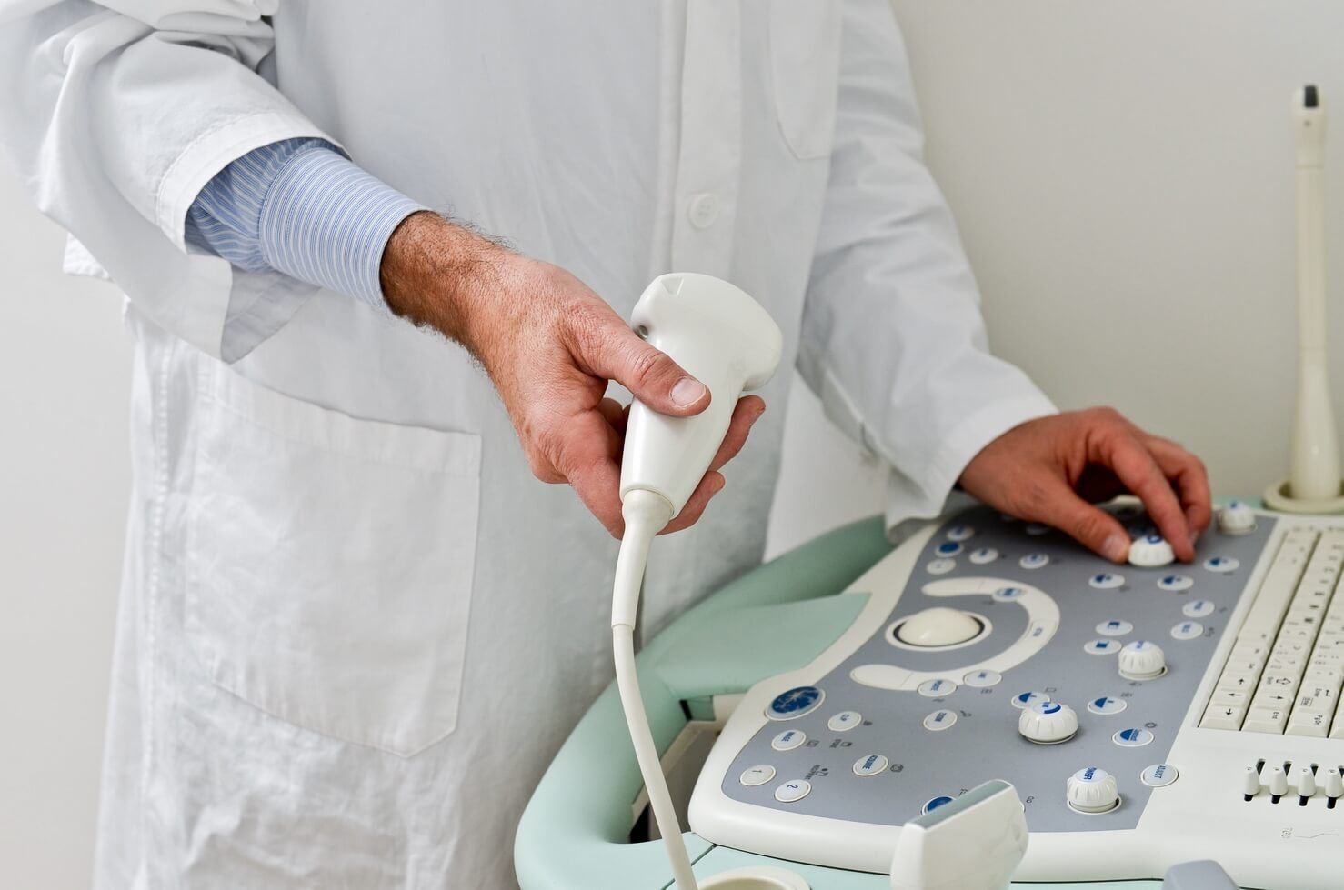
ultrasound examination is a procedure for examining the internal organs of a person, which is based on ultrasound. An ultrasound machine in a small housing contains a sensor and transmitter, which the doctor moves over a person’s skin. The transmitter sends ultrasound into the patient’s body, which is reflected in various ways from different tissues of the internal organs and changes its properties. Catching the reflected ultrasonic waves, the sensor converts them into an electrical signal and sends them to the computer of the ultrasound machine, which converts the arriving signal on the monitor screen into an image. The image on the screen is analyzed by an ultrasound doctor and draws conclusions about the state of the person’s internal organs.
The image on the screen of an ultrasound machine can be 2D, 3D or 4D in color or black and white. 2D is a classic version of ultrasound, and 3D and 4D are experimental image modes that are only available on modern devices. 2D is generally an ordinary flat picture. 3D – three-dimensional image of an internal organ in static state. 4D – three-dimensional image of a human internal organ in motion.
Ultrasound in Nizhny Novgorod is a precise procedure that can detect the disease in its early stages. In addition to all this, ultrasound is absolutely painless and does not pose a danger to human health.
Areas of medicine where ultrasound is used
Ultrasound is used in almost all areas of medicine:
- Cardiology and vascular surgery. Study of blood vessels, heart and veins. Blood flow is examined using Doppler ultrasound. The heart is examined using both a sensor through the sternum and through the esophagus. A 4D echocardiogram makes it possible to obtain a 3D image of the heart in dynamics.
- Gynecology and obstetrics. Ultrasound of the female genital organs, the pregnancy process and the development of the unborn child.
- Internal organs. Search for various abnormalities of human internal organs: pancreas, kidneys, liver, prostate gland, bladder, spleen and others.
- Echoencephalography. Examination of the brain using ultrasound.
- Ophthalmology. Ultrasound is used to determine the location of the lens and measure the size of the eye.
- And others.
What does ultrasound detect?
On the monitor screen of an ultrasound machine, ultrasound shows a two-dimensional, three-dimensional static or dynamic color or black-and-white image of internal organs, blood flow and vessels. The image may depend on modifications to the ultrasound machine itself.
The movement of blood and organ flow is determined through the Doppler effect, when ultrasonic waves are reflected from moving objects with a variable frequency. Changing the ultrasound frequency allows you to understand the nature of the movement of blood flow or the heart. In addition, Doppler ultrasound can be used to construct a 3D image of human organs when the sensor and transmitter move.
A volumetric image of the heart in dynamics makes it possible to obtain a 4D echocardiogram on the monitor screen, which is carried out to study the heart, helping to assess the structure, volume and functioning of this organ.
Based on the patient’s complaints, the doctor selects the correct ultrasound operating modes and the area of application of the sensor with the goal of extremely accurate study of the required organ. The medical conclusion and the correctness of the diagnosis may largely depend on the professionalism of the ultrasound diagnostician. An ultrasound machine is capable of showing pathology even at a very early stage, but the fact how the final results of its activity will be interpreted depends on the competence of the doctor.


